
3.3 Acute Limb Ischaemia – Peripheral Artery Thrombosis
📄 Definition
Acute limb ischaemia (ALI) is a sudden reduction in arterial blood flow to a limb, typically occurring over hours to days, threatening tissue viability and requiring urgent intervention.
⚠️ Causes
🧠 Mnemonic: SAD TOES
Stroke of embolus (e.g. AF, left ventricular thrombus)
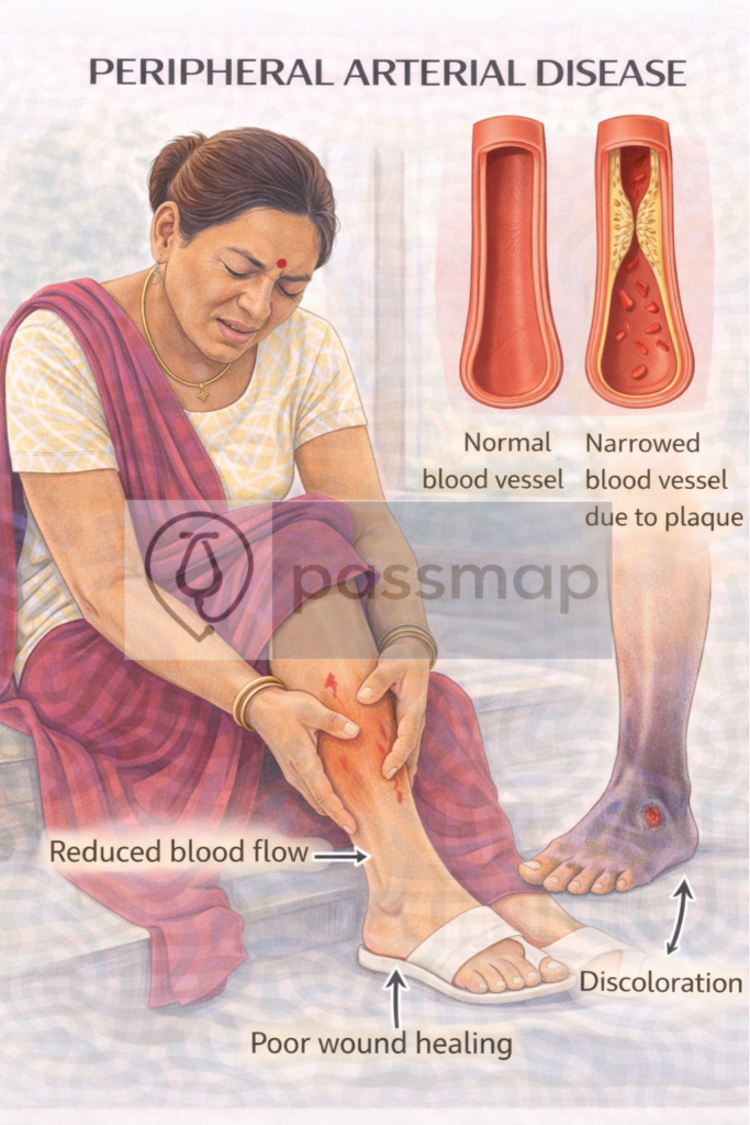
Atherosclerotic plaque rupture and thrombosis
Dissecting aortic aneurysm (rare)
Trauma (arterial injury or occlusion)
Operative complications (e.g. graft occlusion)
External compression (e.g. compartment syndrome)
Spontaneous arterial thrombosis in PAD
6 Ps – Classic Features
Pain (sudden, severe, distal)
Pallor
Pulselessness
Perishing cold (coldness)
Paraesthesia
Paralysis (late sign – poor prognostic factor)
🔺 Sensory loss and paralysis indicate imminent limb loss.
🩺 Clinical Assessment
Capillary refill – delayed
Temperature – compare limbs
Pulse exam – femoral, popliteal, posterior tibial, dorsalis pedis
Buerger’s Test – limb elevation worsens pallor
🧪 Investigations –
🥇 First-Line (Initial Bedside & Emergency)
ECG – Look for AF (most common embolic source)
Doppler Ultrasound (Handheld) – Pulse presence/absence
ABPI – May be unreliable in ALI, but attempted
Bloods: FBC, U&Es, Clotting, Glucose, Lactate, CRP/ESR
Neurovascular Examination – Document baseline function
🥈 Second-Line (Confirmatory Imaging)
CT Angiography (CTA) – Gold standard for vessel occlusion
Duplex Ultrasound (DUS) – If CTA unavailable or contrast contraindicated
MRI Angiography – Less used due to accessibility
🥉 Tertiary / Monitoring
Troponin – Rule out MI in context
Echo – Check for thrombus or valvular embolic source
Thrombophilia screen – In younger or recurrent cases
🚨 Severity – Rutherford Classification
Viable – No immediate threat
Threatened – Urgent intervention needed
Irreversible – Limb non-viable, risk of sepsis
Management
🧠Mnemonic: ACT FAST
Anticoagulate (IV heparin)
Confirm diagnosis (CTA)
Team referral (Vascular surgery)
Fasciotomy (if compartment syndrome risk)
Angioplasty/thrombectomy/embolectomy
Supportive: analgesia, fluids, monitor vitals
Thrombolysis – Consider if no contraindications
📆 Follow up
Long-term antiplatelet (e.g. clopidogrel)
Treat underlying cause (e.g. AF with DOAC)
Smoking cessation, exercise, statin, BP control
Duplex surveillance if stented or revascularised
🔎 Key PARA Exam Traps
💡When answering a question on a cold, pulseless leg, follow this logic:
First Step: Handheld Doppler (to confirm absence of flow).
Immediate Treatment: IV Unfractionated Heparin bolus (to stop the clot from growing).
Gold Standard Image: CT Angiography (to find the blockage).
The “Trap”: If the leg is paralysed and rigid (Rutherford III), the answer is Amputation, not revascularization (due to the risk of death from reperfusion injury).
🔺Last updated in line with:
NICE NG106 (2026 Update): Focusing on standardized pathways for Acute Limb Ischaemia (ALI) and the integration of Handheld Doppler assessment in primary care.
Vascular Society of Great Britain and Ireland (2025): New standards for the “Time to Reperfusion” for Rutherford IIb (Immediately Threatened) limbs.
NICE TA (Technology Appraisals) 2024/25: Regarding the use of mechanical thrombectomy devices in peripheral arterial occlusion.
PARA/MLA-aligned: Fully reviewed February 2026 for the UK Medical Licensing Assessment and Physician Associate Registration Assessment.
🔒 PASSMAP Assurance: All content is peer-reviewed, NICE-compliant, and optimized for the 2026 UK clinical examination cycle.
Educational platform. Not medical advice.