14. Disorders of the Gallbladder & Biliary Tract

(aligned to GMC PARA map — gallbladder is listed, biliary tract folded in for clinical completeness)

📄 Definition

A spectrum of conditions affecting the gallbladder and biliary tract, most caused by gallstones (cholelithiasis) → may lead to pain, infection, obstruction, jaundice, or pancreatitis.

Gallstone types:

  • Cholesterol (80%, linked to 5Fs).

  • Pigment (black/brown; haemolysis, infection).

  • Mixed (commonest in practice).

🛡️ Risk Factors “5 Fs” classic

  • Female, Forty, Fat, Fertile, Fair (Caucasian).

  • ↑ Risk in South Asians (higher pigment stone prevalence in UK).

  • Other: haemolysis (pigment stones), rapid weight loss, diabetes, pregnancy, Crohn’s (ileal disease).

🔬 Pathophysiology of Gallbladder Disorders

DisorderPathophysiology 
Biliary colicGallstone transiently obstructs cystic duct → ↑ pressure → visceral pain. No infection.
Acute cholecystitisPersistent cystic duct obstruction → bile stasis → bacterial infection (E. coli, Klebsiella, Enterococcus) → inflammation + oedema.
EmpyemaUntreated acute cholecystitis → pus in lumen → high risk sepsis → surgical emergency.
Chronic cholecystitisRecurrent inflammation → fibrosis + shrunken gallbladder → dysmotility → ↑ gallbladder cancer risk.
Gallstone ileusLarge stone erodes wall → cholecysto-enteric fistula → stone enters bowel → SBO (terminal ileum).
Gallbladder cancerChronic inflammation (stones, porcelain GB) → dysplasia → adenocarcinoma. Poor prognosis.
 

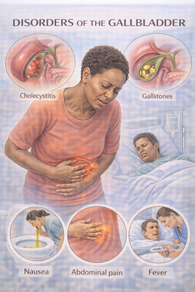
⚠️ Core Gallbladder Conditions

Disorder Key Features Exam Traps
Biliary colic RUQ/epigastric pain (episodic, after fatty meals), radiates to scapula, no fever/jaundice. Pain not truly colicky — constant. Bloods normal.
Acute cholecystitis RUQ pain + fever, Murphy’s sign +, raised WCC/CRP. USS: wall thickening + pericholecystic fluid.
Empyema of GB Pus-filled gallbladder, septic patient. Needs urgent IV abx + drainage.
Chronic cholecystitis Recurrent low-grade RUQ pain, fibrosis. Follows repeated acute attacks.
Gallstone ileus Small Bowel Obstruction (SBO) from stone via fistula. Elderly women. Rigler’s triad on AXR/CT: SBO + pneumobilia + ectopic stone.
Gallbladder cancer Rare, linked to chronic stones/porcelain GB. Poor prognosis, often incidental.

📄 Jaundice Framework — Pre-hepatic, Hepatic, Post-hepatic

TypePathophysiologyKey FeaturesTypical CausesTest Clues
Pre-hepatic↑ Haemolysis → excess unconjugated bilirubin exceeds liver conjugation capacityMild jaundice, no bilirubinuria, dark stoolsHaemolysis (AIHA, sickle cell, G6PD, malaria)↑ unconjugated bilirubin, normal LFTs, anaemia, ↑ retics
HepaticHepatocellular injury or impaired conjugation/excretion → mixed hyperbilirubinaemiaJaundice ± systemic illness, CLD stigmataViral hepatitis, ALD, MASLD, cirrhosis, drugs↑ ALT/AST > ALP, deranged INR/albumin
Post-hepatic (obstructive)Blocked bile ducts → conjugated bilirubin refluxes into bloodDark urine, pale stools, pruritus, RUQ painGallstones, strictures, cholangiocarcinoma, pancreatic cancer↑ conjugated bilirubin; ↑ ALP/GGT > ALT; duct dilatation on US/MRCP

💡 Exam Hotspot – Gilbert’s Syndrome

  • Definition: Benign inherited ↓ UDP-glucuronyl transferase → impaired conjugation of bilirubin.

  • Features: Isolated unconjugated hyperbilirubinaemia; intermittent mild jaundice (triggered by fasting, illness, alcohol, stress).

  • Tests: Normal LFTs, no haemolysis, only ↑ unconjugated bilirubin.

  • Exam Trap: Don’t confuse with hepatic pathology — everything else is normal.

  • Management: Reassure — no treatment required.

  • Placement note: Not a GMC PARA core condition, but very common in exam vignettes to test your jaundice framework knowledge.

🧠 Exam triggers:

  • Dark urine + pale stools + pruritus → obstructive/ post-hepatic

  • Normal LFTs + unconjugated bilirubin → pre-hepatic/ Guilbert’s (benign)

  • Stigmata of CLD → hepatic.

  • Anaemia + ↑ reticulocytes + jaundice → pre-hepatic haemolysis.

📎 See also: “Liver Failure (including Cirrhosis)” for detailed intra-hepatic causes and management.

⚠️ Core Biliary Tract Conditions

DisorderKey FeaturesExam Traps
CholedocholithiasisStone in common bile duct (CBD) → RUQ pain, cholestatic LFTs (↑ALP, ↑bilirubin).USS may miss CBD stones → MRCP better.
Ascending cholangitisInfection + obstruction → Charcot’s triad: RUQ pain, fever, jaundice. Reynolds’ pentad if + hypotension & confusion (sepsis).Always needs IV abx + urgent biliary decompression (ERCP).
Obstructive jaundicePainless jaundice (think malignancy: pancreatic/ampullary/CCA) vs painful (stones).Courvoisier’s law: palpable, non-tender GB unlikely due to stones (think cancer).
Gallstone pancreatitisGallstone impacts ampulla → blocks bile + pancreatic ducts → acute pancreatitis (lipase > amylase).Do not confuse with biliary colic/cholecystitis. USS first-line, MRCP for CBD stones. Cholecystectomy after recovery to prevent recurrence.
 

🔬 Pathophysiology of Biliary Tract Disorders

DisorderPathophysiology
CholedocholithiasisGallstone migrates from gallbladder → lodges in common bile duct (CBD) → obstruction of bile outflow → conjugated hyperbilirubinaemia, cholestatic LFTs.
Ascending cholangitisObstructed CBD becomes infected (E. coli, Klebsiella, Enterococcus) → ↑ pressure + bacterial proliferation → systemic sepsis.
Obstructive jaundice (stones/malignancy)Bile cannot drain → conjugated bilirubin refluxes into blood → dark urine, pale stools, pruritus. Chronic obstruction → secondary biliary cirrhosis.
Gallstone pancreatitis Stone at ampulla of Vater blocks both CBD + pancreatic duct → reflux/activation of pancreatic enzymes → acute pancreatitis.
🧠 Key exam points:
  • Choledocholithiasis = obstruction, no infection.

  • Cholangitis = obstruction + infection (Charcot’s triad ± Reynolds’ pentad).

  • Obstructive jaundice can be benign (stone, stricture) or malignant (pancreatic/ampullary/CCA).

  • Gallstone pancreatitis is a biliary complication but often tested under pancreas.

📦 Additional: Autoimmune Cholestatic Disorders (Not PARA Core, but Exam-Linked)

FeaturePrimary Biliary Cholangitis (PBC)Primary Sclerosing Cholangitis (PSC)
WhoMiddle-aged women (F>M)Young/middle-aged men (M>F), strong IBD (UC) link
PathophysiologyAutoimmune destruction of intra-hepatic bile ductsInflammation + fibrosis of intra & extra-hepatic bile ducts (“onion-skin”)
ClinicalPruritus, fatigue, jaundice; xanthelasma/xanthomas, osteoporosisFatigue, pruritus, jaundice; recurrent cholangitis; progression to cirrhosis
AntibodyAMA (anti-mitochondrial, 90–95%)p-ANCA (non-specific)
LFTsCholestatic: ↑ALP, ↑GGT > ↑ALT/ASTCholestatic: ↑ALP, ↑GGT; may be fluctuating
ComplicationsCirrhosis, portal HTN, ↑ risk hepatocellular carcinomaCirrhosis, cholangiocarcinoma (10–15%), colorectal cancer (with UC)
DiagnosisAMA + cholestatic LFTs ± liver biopsyMRCP/ERCP: multifocal strictures + dilatations (“beading”)
ManagementUDCA (ursodeoxycholic acid), symptom relief (cholestyramine for itch), liver transplant if end-stageERCP for dominant strictures, manage complications, liver transplant if end-stage

🧠 Memory hook

  • PBC = AMA, intra-hepatic, women, xanthelasma

  • PSC = p-ANCA, beading ducts, men with UC, cholangiocarcinoma risk

🔬 Investigations 

StepInvestigationPurpose
1️⃣ InitialSerum amylase or lipase (>3× ULN)Diagnostic – lipase preferred (more sensitive/specific).
 FBC, CRP, U&E, LFTs, glucose, calcium, ABGAssess systemic impact, inflammation, severity.
2️⃣ ImagingUSS abdomen (first-line)Detect gallstones, gallbladder wall thickening, duct dilatation.
 MRCPNon-invasive biliary tree assessment – choledocholithiasis, obstruction.
 ERCPDiagnostic + therapeutic (stone removal, stenting) – not first-line.
 CT abdomen (contrast)If complications suspected (perforation, pancreatitis, ileus, cancer).
3️⃣ Prognostic (if pancreatitis suspected)Glasgow-Imrie score, CRP >150, APACHE IIPredicts severity, guides HDU/ICU

💊 Management (Stepwise – NICE / BSG)

ConditionFirst-lineEscalation / Notes
Biliary colicAnalgesia (NSAIDs/opioids), lifestyle adviceElective laparoscopic cholecystectomy if recurrent
Acute cholecystitisAdmit, IV fluids, analgesia, IV antibiotics (co-amoxiclav)Laparoscopic cholecystectomy <1 week (ideally within 72 hrs)
CholedocholithiasisERCP stone extraction ± stentThen laparoscopic cholecystectomy
CholangitisIV antibiotics (pip-taz or ceftriaxone + metronidazole), sepsis 6Urgent ERCP for biliary drainage
Gallbladder empyemaIV antibiotics + drainageSurgical consult urgent
Gallbladder cancerStaging CT, MDT discussionSurgery ± palliative care

📋 Follow-Up

  • Post-cholecystectomy: recovery usually complete; warn about occasional bile salt diarrhoea.

  • Gallstone pancreatitis: offer cholecystectomy during same admission.

  • Gallbladder cancer: oncology/hepatobiliary follow-up, often palliative.

  • PSC/PBC (if overlap suspected): hepatology monitoring.

🔎 Key PARA Exam Traps

  • Murphy’s sign = cholecystitis, not biliary colic.

  • Charcot’s triad ± Reynold’s pentad = cholangitis (sepsis, urgent ERCP).

  • ERCP = therapeutic, not just diagnostic.

  • Asymptomatic stones = no treatment unless porcelain GB, sickle cell, or very high risk.

  • Courvoisier’s law = malignancy > stones.

  • USS first-line, MRCP next for ductal stones, ERCP = therapeutic.

  • Biliary colic = pain only, normal bloods.

  • Acute cholecystitis = pain + fever + raised WCC/CRP.

  • Lipase > amylase for diagnosing gallstone pancreatitis.

📅 Last updated in line with NICE NG104 (Gallstone disease) & BSG Guidelines

Published: 2014 • Last updated: 2022 • Reviewed: August 2025
🔒 PASSMAP ensures all content is PARA-aligned, exam-focused, and NICE-compliant.

Scroll to Top